How Dentists Diagnose the Need for Periodontal Treatment
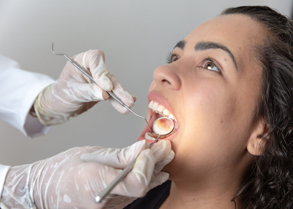
Most people assume gum disease is easy to spot. If the gums bleed, there is a problem. If they do not, everything must be fine. In practice, it is rarely that simple. Some patients with advanced periodontal breakdown report almost no pain. Others come in worried about minor bleeding that turns out to be reversible gingivitis. The dentist’s job is to sort those situations carefully, using a combination of history, visual examination, measurements, imaging, and clinical judgment.
That process matters because periodontal disease is not just about irritated gums. It is an infection and inflammatory condition that affects the structures supporting the teeth, including gum tissue, bone, and periodontal ligament. Once those supporting tissues are lost, they do not simply grow back on their own. Early diagnosis can make the difference between a straightforward deep cleaning and a much longer course of Periodontal Treatment that may involve surgery, maintenance visits, and strict home care over years.
A thorough diagnosis is never based on one sign alone. It is built from a pattern.
The first clues often appear before the examination starts
Before a dentist or hygienist even picks up an instrument, useful diagnostic information starts coming in. The patient may describe bleeding when brushing, bad breath that does not improve with mouthwash, tenderness in one area, gum recession, loose teeth, or food packing between teeth that used to feel tight. Sometimes the concern comes from cosmetic changes rather than discomfort. A patient may say, “My teeth look longer,” or “My smile seems uneven now.” Those are classic observations when gum recession has begun.
Medical history also shapes the suspicion level. Diabetes, especially when poorly controlled, often changes how the gums respond to plaque and how quickly breakdown progresses. Smoking is another major factor. It can suppress obvious signs like bleeding, which means the gums may look less inflamed than they truly are. Pregnancy, certain medications, dry mouth, immune conditions, and a history of previous periodontal therapy all influence what the dentist expects to find.
Age plays a role, but not in the simplistic way many people think. Severe periodontal disease is more common in adults because tissue destruction is cumulative, but younger patients can absolutely develop significant disease, especially if there is a strong genetic tendency, smoking, uncontrolled systemic disease, or long periods without professional care. I have seen patients in their early thirties who were shocked to learn they had pockets measuring 6 or 7 millimeters around molars, despite brushing daily. Habit alone does not tell the full story. Technique, anatomy, risk factors, and the body’s inflammatory response all matter.
What the dentist looks for during the visual exam
The visual exam gives an immediate sense of gum health. Healthy gums are usually firm, well adapted around the teeth, and generally coral pink, though natural pigmentation varies. Diseased gums may appear swollen, shiny, red, dusky, or uneven. In some patients, the tissue margin looks rolled rather than knife edged. In others, the gums have pulled away, exposing root surfaces and creating the appearance of longer teeth.
Plaque and tartar are central findings. Plaque is the soft bacterial film that forms constantly. Tartar, also called calculus, is plaque that has hardened. Tartar above the gumline is easy for patients to see sometimes, but tartar below the gumline is more important from a periodontal perspective because it holds bacteria against the root and keeps inflammation active. A patient may feel that their teeth are smooth enough, while subgingival deposits remain firmly attached in deeper pockets.
Dentists also watch for bleeding, either spontaneous or triggered gently during the exam. Bleeding is a sign of inflamed tissue, but its absence does not automatically rule out disease. Smokers are the classic example. Their tissues may bleed less than expected because nicotine affects blood flow. That is one reason no careful clinician diagnoses or clears a patient based only on whether the gums bleed during brushing.
Tooth position can offer more clues. Drifting teeth, spaces that were not there before, and changes in bite may suggest loss of periodontal support. In advanced cases, front teeth begin to flare outward or separate slightly. Patients often notice the cosmetic effect before they understand the cause. Mobility is another important sign. A tooth with reduced bone support may move slightly when tested. That movement can also come from biting forces, recent trauma, or infection around the tip of the root, so the dentist has to distinguish among those possibilities.
Periodontal probing is one of the most important diagnostic tools
If there is one part of the exam that determines whether Periodontal Treatment is needed, it is usually periodontal probing. This is where the dentist or hygienist uses a thin measuring instrument, called a periodontal probe, to check the depth of the space between the tooth and the gum.
In health, that space is shallow. A reading of 1 to 3 millimeters is generally considered within normal limits when there is no bleeding, recession, or other pathology. Once the probe records deeper measurements, especially 4 millimeters and beyond with bleeding or radiographic bone loss, concern rises. Deeper readings suggest the gum attachment has migrated downward and a periodontal pocket has formed.
A full periodontal chart records six measurements per tooth. That level of detail matters because gum disease is not always uniform. A patient may have relatively healthy front teeth and much deeper pockets around molars, especially on the tongue side where cleaning is harder. Without a full chart, those site-specific problems are easy to miss.
Bleeding on probing adds another layer of information. It tells the clinician that the tissue lining the pocket is inflamed and fragile. Suppuration, or pus, is a stronger sign of active infection. Recession must also be included in the picture. A tooth may have a shallow probing depth because the gum has already receded significantly, while the total attachment loss is still substantial. That is why dentists look beyond pocket depth alone and assess clinical attachment level, which combines probing depth with the position of the gum margin.
There is nuance here. A single 4 millimeter reading does not automatically mean severe periodontal disease. Inflamed gums from reversible gingivitis can create pseudo pockets, where swelling increases the measurement without true attachment loss. That is why probing must be interpreted alongside the rest of the exam. Repeated measurements over time are often revealing. If a site remains deep after inflammation is reduced, or if bone loss is visible on X-rays, the case for periodontal disease becomes much stronger.
X-rays show what the eye cannot
Gums can hide a great deal. X-rays are essential because they reveal the level and pattern of supporting bone around the teeth. A dentist diagnosing periodontal disease pays close attention to how high the bone sits relative to the roots, whether the loss is generalized or localized, and whether it appears horizontal or vertical.
Horizontal bone loss is more even across several teeth. Vertical bone loss creates angular defects that can be more complex to treat. Furcation involvement, where bone loss affects the area between the roots of molars, is another important finding. Molars with furcation involvement are harder to clean, often harder to stabilize, and frequently require more advanced periodontal care.
Radiographs also help rule out look-alike problems. A tooth can be sore or loose because of an abscess at the root tip, a vertical root fracture, or trauma from occlusion. Those conditions may overlap with periodontal issues or mimic them. A clear diagnosis often depends on combining X-ray findings with probing, mobility, and symptoms.
One important limitation deserves mention. X-rays do not show active inflammation directly. They show past destruction of hard tissue. A patient may have severe bleeding and deep pockets before major bone loss becomes obvious on imaging. Conversely, old bone loss may be present in a stable patient whose disease is no longer actively progressing. That is why no responsible dentist diagnoses the need for Periodontal Treatment from X-rays alone.
The difference between gingivitis and periodontitis
This distinction sits at the center of diagnosis. Gingivitis is inflammation limited to the gums. It causes redness, swelling, and bleeding, but no loss of bone or connective tissue attachment. Periodontitis goes further. It involves irreversible destruction of the structures that support the teeth.
At chairside, the difference is determined by evidence of attachment loss. If the gums are inflamed but the bone levels are normal and the probing findings can be explained by swelling alone, the diagnosis leans toward gingivitis. If there are deeper pockets, recession, radiographic bone loss, furcation involvement, or mobility related to loss of support, periodontitis becomes the concern.
This matters because treatment plans differ. Gingivitis often improves with a professional cleaning and stronger daily plaque control. Periodontitis usually requires more than that, often including scaling and root planing, targeted reevaluation, and sometimes referral to a periodontist. Patients are often surprised when they are told they need something more involved than a standard cleaning. From their perspective, they came in for routine care. From the clinician’s perspective, the measurements and X-rays show a chronic disease process that a routine prophylaxis will not resolve.
Why symptoms do not always match severity
One of the more frustrating aspects of periodontal diagnosis is how quiet the disease can be. Unlike a toothache, which can be sharp and impossible to ignore, periodontal disease often progresses with very little pain. Patients may adapt gradually to bleeding, mild bad breath, or occasional tenderness and stop noticing it. Others interpret gum recession as normal aging and do not realize it can reflect tissue breakdown.
I have heard many versions of the same sentence in clinical settings: “If it were serious, I would have felt something.” Unfortunately, that belief leads many people to delay care. By the time they notice tooth mobility or visible shifting, significant support may already be gone.
There are also patients at the opposite end https://danteusmz638.wpsuo.com/periodontal-treatment-for-chronic-gingivitis-and-periodontitis of the spectrum, highly alert to every sign. A little bleeding after flossing can make them fear major disease. Sometimes that bleeding comes from inconsistent flossing or a rough brushing technique rather than established periodontitis. Good diagnosis protects both groups, the patients who minimize symptoms and the patients who overread them.
Risk assessment changes the threshold for concern
Diagnosis is not only about what the tissues look like today. It is also about how likely the condition is to worsen. Two patients can present with similar probing depths and very different risk profiles.
A nonsmoker with excellent diabetes control, mild localized bone loss, and strong home care may respond well to conservative treatment and careful monitoring. A heavy smoker with generalized 5 to 6 millimeter pockets, poor plaque control, and long gaps between dental visits has a very different outlook. The disease in the second patient is more likely to progress and less likely to heal predictably without intensive intervention.
Dentists typically pay close attention to factors such as smoking, diabetes, family history, stress, bruxism, and oral hygiene habits. They also consider restorative issues. Crowded teeth, overhanging fillings, open contacts, and poorly contoured crowns can trap plaque and create localized periodontal problems. Wisdom tooth anatomy and deep grooves on root surfaces can make some sites stubbornly difficult to maintain, even in motivated patients.
This is where real clinical judgment comes in. The same 4 millimeter pocket may be monitored in one patient and treated more aggressively in another, depending on bleeding, bone loss, systemic factors, and evidence of progression.
When a standard cleaning is not enough
Many patients understandably view all dental cleanings as basically the same. They are not. A routine cleaning is designed to remove plaque and calculus above the gumline and slightly below it in a mouth that is otherwise periodontally healthy. It is preventive care.
Periodontal treatment addresses disease below the gumline. If there are deeper pockets and root surfaces contaminated with bacterial deposits and calculus, a simple cleaning will not reach adequately into those areas. In those cases, scaling and root planing is usually recommended. This deeper cleaning smooths root surfaces and disrupts bacterial biofilm in the pockets. Local anesthetic is often used because the treatment goes into areas that would be uncomfortable otherwise.
Dentists arrive at that recommendation because the diagnostic pieces line up: pocketing, bleeding, attachment loss, bone changes, and clinical signs of active inflammation. Insurance terminology sometimes confuses patients here. They may hear that their cleaning has been “upgraded” and think the office is changing the plan for billing reasons. A sound diagnosis should always be explainable in clinical terms, not administrative ones. A good dentist can show the measurements, point out the X-ray findings, and explain why the tissues need more than a routine polish.
Reevaluation is part of diagnosis, not an afterthought
Periodontal diagnosis is not a one-visit event. After initial therapy, the tissues need time to respond. Rechecking pocket depths, bleeding points, and inflammation after scaling and root planing is essential. Some sites improve dramatically once bacterial deposits are removed and home care improves. Others stay deep and continue to bleed, which may suggest persistent calculus, difficult anatomy, root grooves, furcation involvement, or the need for surgical access.
This reevaluation step often changes the treatment plan. A patient may begin with nonsurgical therapy and then move to periodontal surgery for a few isolated areas that did not respond. Another may stabilize well and shift into periodontal maintenance, with cleanings every three or four months rather than twice a year.
That ongoing monitoring is one reason periodontal care tends to feel different from routine dentistry. It is chronic disease management. The dentist is not only asking, “What do I see today?” but also, “How has this changed over time, and how is this patient responding to treatment?”
Cases that require a closer look
Some situations make diagnosis more complex. A cracked tooth can create an isolated deep pocket that mimics periodontal disease. Endodontic infections can drain through the gum and look periodontal at first glance. Aggressive tissue loss around a single tooth may point to a root fracture, foreign body, or unusual local factor rather than generalized periodontitis.
Orthodontic history also matters. Teeth moved outside the bony housing can develop recession, especially if brushing is forceful and the gum tissue is thin. That recession may not reflect classic periodontal disease, but it still requires careful assessment. Likewise, enlarged gums caused by medication can create deeper measurements that look alarming until the underlying cause is understood.
This is why a good periodontal diagnosis is rarely rushed. The dentist must separate true attachment loss from swelling, local trauma, endodontic lesions, and anatomical variation. It takes experience to know when a deep pocket is “just inflamed” and when it reflects meaningful structural damage.
What patients should expect during a periodontal evaluation
A proper periodontal evaluation should feel methodical. The clinician should review symptoms and health history, inspect the gums visually, take or review appropriate X-rays, probe around the teeth, and discuss the findings in plain language. Patients should hear more than “your gums are bad.” They should be told where the pocketing is, whether bone loss is present, whether the issue is localized or generalized, and what level of Periodontal Treatment is being recommended.
Useful questions from patients often include asking whether the condition is gingivitis or periodontitis, how deep the worst pockets are, whether bone loss is visible, and what improvement is realistic after treatment. Another smart question is whether any contributing factors, such as smoking, diabetes, grinding, or difficult restorations, are making the condition worse.
When the explanation is clear, treatment tends to make more sense. Patients are far more likely to accept therapy when they understand that periodontal disease is not simply “dirty teeth,” but a measurable, progressive breakdown of support around the teeth.
The real goal of diagnosis
Dentists diagnose the need for periodontal treatment to answer a practical question: what does this patient need now to keep teeth healthy and functional for the long term? Sometimes the answer is better home care and routine maintenance. Sometimes it is deep cleaning. Sometimes it is referral to a periodontist for surgery or management of advanced bone loss.
The most important point is that this decision should come from evidence, not guesswork. Probing depths, bleeding, gum appearance, recession, mobility, X-rays, risk factors, and changes over time all contribute to the picture. No single finding stands alone.
When done well, periodontal diagnosis catches disease before teeth become loose, painful, or hopeless. It gives the patient a chance to intervene while treatment is still manageable. That is the value of a careful exam. It turns gum disease from something hidden and progressive into something measurable, explainable, and treatable.
Avra Dental
Address: 1708 S Victoria Ave B, Ventura, CA 93003
Phone number: (805) 941-1001
FAQ About Periodontal Treatment
Can a dentist get rid of periodontal disease?
A dentist or gum specialist (periodontist) cannot fully cure or reverse advanced periodontal disease (periodontitis), but they can successfully stop its progression and manage the infection.
Is periodontitis very serious?
Yes, periodontitis is a very serious, advanced form of gum disease that destroys the bone and tissues supporting your teeth.
How is stage 2 periodontal disease treated?
Stage 2 periodontal disease (early to moderate periodontitis) is primarily treated with non-surgical deep cleaning procedures like scaling and root planing to remove bacteria and tartar below the gumline.